Raman microscopy is a powerful analytical technique that allows researchers to identify and analyze microplastics, even those as small as 1 μm in diameter. By combining Raman spectroscopy and optical microscopy, it provides a unique molecular fingerprint of the sample, enabling the distinction between different types of polymers, contaminants, and dyes.
Key Benefits of Raman Microscopy for Microplastic Analysis:
- High spatial resolution for examining tiny particles
- Minimal sample preparation and ability to analyze wet samples
- Detailed information on the chemical composition of microplastics
Microplastics are small plastic particles less than 5mm in size, originating from various sources such as synthetic textiles, vehicle tires, personal care products, and city dust. Identifying microplastics is crucial for understanding their impact on the environment and human health, as they have been found in marine organisms, seafood, and even drinking water.
Steps in Raman Microscopy for Microplastic Analysis:
-
Sample Collection and Preparation
- Water, soil, and air samples are collected using specialized equipment
- Samples are filtered, washed, dried, and separated from other particles
-
Raman Microscope Setup
- Calibration and configuration of the microscope and software
- Proper alignment of the laser and sample holder
-
Obtaining Raman Spectra
- Laser excites the molecules, causing them to vibrate and scatter light
- Scattered light is collected and analyzed to produce a unique Raman spectrum
-
Identifying Microplastics
- Raman spectra are compared to reference libraries of known microplastic materials
- Peak positions, intensities, and shapes are matched to determine the type of microplastic
By leveraging Raman microscopy, researchers can accurately identify and characterize microplastics, enabling targeted strategies to mitigate their impact on the environment and human health.
Sources and Types of Microplastics
Microplastics are small plastic particles less than 5mm in size. They can enter the environment directly (primary microplastics) or break down from larger plastics (secondary microplastics). The International Union for Conservation of Nature (IUCN) identifies seven primary sources of microplastics in the marine environment:
Primary Sources of Microplastics
| Source | Description |
|---|---|
| Synthetic textiles | Microfibres from clothing and other synthetic materials |
| Vehicle tyres | Tyre wear generates microplastics, especially from electric vehicles |
| Road markings | Paint and other materials used on roads can contain microplastics |
| Personal care products and cosmetics | Some products contain microbeads or other microplastics |
| Plastic pellets | Small plastic pellets used in manufacturing can enter the environment |
| Marine coatings | Paints and coatings used on boats and ships can contain microplastics |
| City dust | Artificial turf, building paints, and industrial abrasives can generate microplastics |
Microfibres from textiles are a significant contributor to micropollution. Although only synthetic microfibres are considered microplastics, micro fragments from all types of fibres, including natural ones like cotton and wool, also contribute to pollution.
Vehicle tyres are another significant source of microplastics, with an estimated 0.5 million metric tonnes generated from tyre wear in the European Union per year.
City dust, which includes artificial turf, building paints, and industrial abrasives, is also a significant source of microplastics. Artificial turf, for example, contains polymeric infill made from recycled vehicle tyres, which can be accidentally removed during maintenance or use.
Understanding the sources and types of microplastics is crucial in addressing microplastic pollution. By identifying the primary sources of microplastics, we can develop targeted strategies to reduce their impact on the environment.
Why Identify Microplastics?
Microplastics have become a significant environmental issue, and identifying them is crucial for understanding their impact on the environment and human health. Microplastics have been found in marine organisms, commercial seafood, and even drinking water. Standard water treatment facilities cannot remove all traces of microplastics.
The Importance of Identification
Identifying microplastics is essential for:
- Developing effective strategies to reduce their impact on the environment
- Understanding their effects on human health
- Creating targeted solutions to reduce their release into the environment
For instance, identifying microplastics in personal care products and cosmetics can lead to the development of alternative products that do not contain microbeads.
Human Health Impacts
Experts estimate that humans consume microplastics in the range of 74,000 to 121,000 particles per year through ingestion and inhalation. The health effects of this consumption on humans are still unknown, and identifying microplastics is the first step towards understanding these effects and developing strategies to mitigate them.
In conclusion, identifying microplastics is a critical step towards addressing microplastic pollution. It enables us to understand their sources, effects on the environment and human health, and develop targeted strategies to reduce their impact.
Raman Microscopy for Microplastic Analysis
Raman microscopy is a powerful tool for identifying microplastics. It offers high precision and can analyze very small particles, even those as small as 1 μm in diameter. This technique combines Raman spectroscopy and optical microscopy to examine microscopic pieces of plastics.
How Raman Microscopy Works
Raman microscopy focuses a laser beam onto a small spot to obtain Raman spectra. This allows researchers to analyze the chemical composition of microplastics. The high spatial resolution of Raman microscopy enables the examination of even the smallest microplastic particles, which is crucial when researching the impacts of plastic on human health.
Advantages of Raman Microscopy
Raman microscopy has several advantages over other analytical techniques:
- High spatial resolution: Enables the examination of small microplastic particles
- Minimal background interference: Ideal for analyzing wet samples
- Detailed information: Provides information about the chemical composition of microplastics
By using Raman microscopy, researchers can gain valuable insights into the characteristics of microplastics, ultimately informing strategies to mitigate their effects.
How Raman Spectroscopy Works
Raman spectroscopy is a non-invasive analytical technique that uses light to identify the chemical composition of microplastics. Here's how it works:
The Process
1. A laser beam is focused onto a small spot, exciting the molecules and causing them to vibrate.
2. These molecular vibrations lead to a change in the frequency of the scattered light.
3. The resulting Raman spectrum is a unique fingerprint of the molecular structure, allowing researchers to identify the chemical composition of the microplastic.
Advantages of Raman Spectroscopy
| Advantage | Description |
|---|---|
| High spatial resolution | Enables the examination of microplastic particles as small as 1 μm in diameter |
| Minimal background interference | Ideal for analyzing wet samples |
| Detailed information | Provides information about the chemical composition of microplastics |
By using Raman spectroscopy, researchers can gain valuable insights into the characteristics of microplastics, ultimately informing strategies to mitigate their effects on the environment and human health.
Key Features of Raman Microscopy
Raman microscopy is a powerful tool for identifying microplastics, offering several key features that make it an ideal technique for this application.
High Spatial Resolution
Raman microscopy allows for the examination of microplastic particles as small as 1 μm in diameter. This high spatial resolution is essential for studying microplastics, as they can be absorbed into the human gut and have potential health implications.
Minimal Background Interference
Raman spectroscopy is ideal for analyzing wet samples, with minimal water interference. This is in contrast to other techniques, such as Fourier Transmission Infra-Red Spectroscopy (FTIR), which can be affected by water interference. Additionally, Raman microscopy can reduce background fluorescence, allowing for more accurate analysis of microplastics.
Detailed Information
Raman spectroscopy provides detailed information about the chemical composition of microplastics, allowing researchers to identify the type of polymer present. This information is crucial for understanding the sources and fate of microplastics in the environment and their potential impact on human health.
Key Features of Raman Microscopy
| Feature | Description |
|---|---|
| High Spatial Resolution | Examines microplastic particles as small as 1 μm in diameter |
| Minimal Background Interference | Ideal for analyzing wet samples with minimal water interference |
| Detailed Information | Provides information about the chemical composition of microplastics |
By leveraging these key features, Raman microscopy has become a vital tool for identifying and analyzing microplastics, enabling researchers to gain valuable insights into the characteristics of these tiny particles.
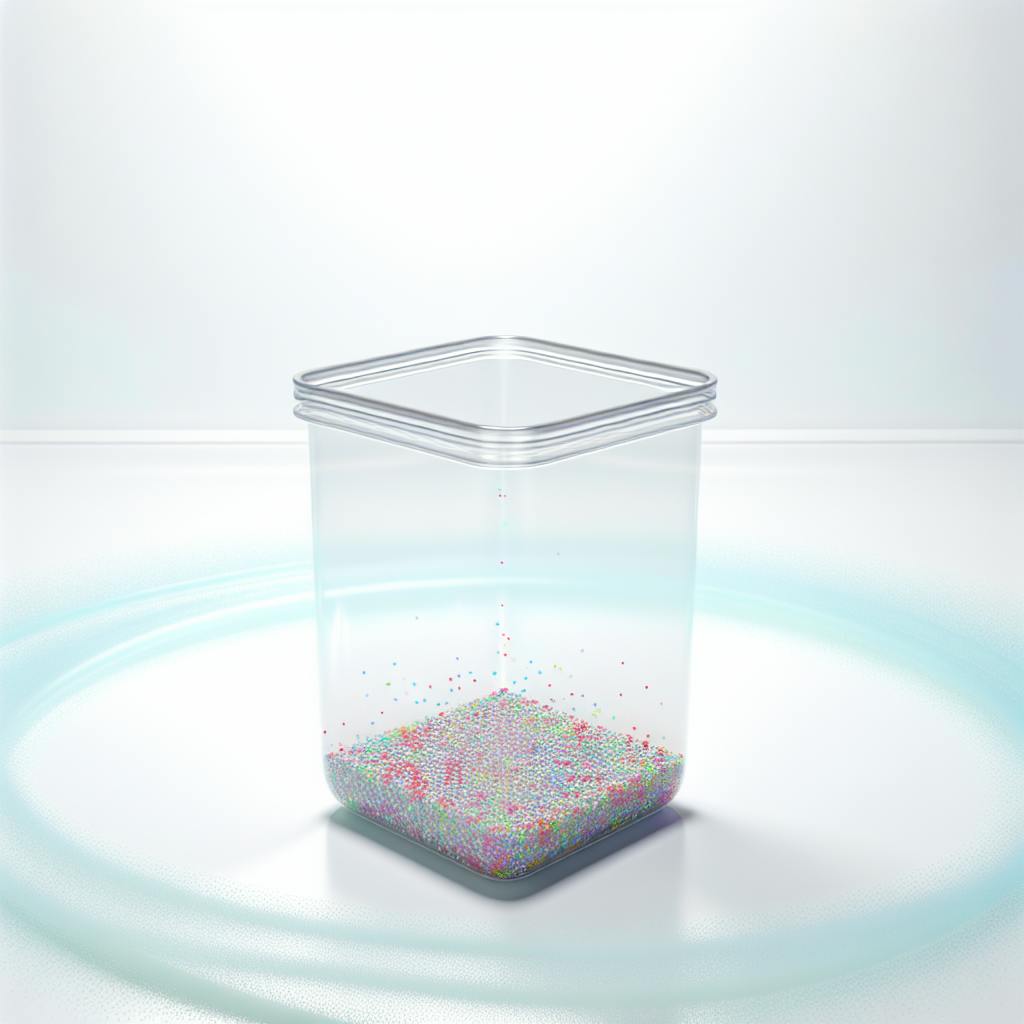
Collecting Microplastic Samples
Collecting microplastic samples is a crucial step in understanding the extent of microplastic pollution in various environments. Microplastics can be found in water, soil, and air, and collecting samples from these sources requires different methods.
Water Samples
To collect microplastic samples from water, researchers use specialized equipment such as:
| Equipment | Description |
|---|---|
| Plankton nets | Capture small particles, including microplastics, from the water surface |
| Sediment corers | Collect samples from the seafloor or lakebed |
| Bottles or containers | Collect water samples, which are then filtered to separate microplastics from other particles |
Soil Samples
Collecting microplastic samples from soil involves:
| Method | Description |
|---|---|
| Digging and collecting soil cores | Collect soil samples from different depths |
| Scooping surface soil samples | Collect soil samples from the surface |
Air Samples
Collecting microplastic samples from air involves using air samplers, which capture particles suspended in the air. Air samplers can be deployed in various locations, including urban and rural areas.
Sample Preparation
Regardless of the sampling method, microplastic samples require careful preparation to ensure accurate analysis. This includes:
- Washing samples to remove contaminants
- Drying samples to prevent degradation
- Separating microplastics from other particles using techniques such as density separation or sieving
By collecting and analyzing microplastic samples from various environments, researchers can gain a better understanding of the sources, fate, and impact of microplastics on the environment and human health.
Preparing Samples for Raman Analysis
Preparing microplastic samples for Raman analysis is a crucial step in obtaining accurate and reliable results. The goal of sample preparation is to ensure that the samples are free from contaminants, dry, and separated from other particles, allowing for optimal Raman analysis.
Filtering and Washing
Collected microplastic samples are first filtered onto silicon nitride nanomembranes using water. This process helps to remove any contaminants and separate the microplastics from other particles. The samples are then washed to remove any remaining impurities.
Drying
After filtering and washing, the samples are dried to prevent degradation and ensure that the Raman analysis is not affected by moisture. Drying can be done using various methods, including air drying or using a desiccator.
Separating Microplastics from Other Particles
To separate microplastics from other particles, techniques such as density separation or sieving can be used. The following table outlines the separation methods:
| Separation Method | Description |
|---|---|
| Density Separation | Uses a density gradient to separate particles based on their density |
| Sieving | Uses a mesh to separate particles based on their size |
Mounting Samples
Once the samples are prepared, they are mounted onto a microscope slide or a specialized Raman microscope sample holder. This ensures that the samples are securely held in place during the Raman analysis.
By following these steps, microplastic samples can be prepared for optimal Raman analysis, allowing researchers to accurately identify and characterize microplastics.
Setting Up the Raman Microscope
To analyze microplastics using a Raman microscope, you need to set it up correctly. This section will guide you through the process.
Calibration
Calibration is essential to ensure accurate results. You need to adjust the microscope's laser and spectrometer using a reference material, such as silicon. This ensures that the Raman peaks are correctly aligned.
Configuration
Once calibrated, configure the microscope's software to collect Raman spectra from microplastic samples. Set the software to collect spectra at specific wavelengths and laser powers for optimal results.
Sample Holder
The sample holder is a crucial component. It holds the microplastic sample in place during analysis, ensuring the sample is securely positioned. Design the sample holder to accommodate the specific type of microplastic sample being analyzed.
Laser Alignment
Laser alignment is critical. Align the laser to focus on the microplastic sample by adjusting the microscope's mirrors and lenses.
By following these steps, you can ensure your Raman microscope is properly set up for microplastic analysis.
Setup Checklist
| Step | Description |
|---|---|
| Calibration | Adjust laser and spectrometer using a reference material |
| Configuration | Set software to collect Raman spectra at specific wavelengths and laser powers |
| Sample Holder | Design holder to accommodate specific microplastic sample type |
| Laser Alignment | Align laser to focus on microplastic sample |
Remember to follow these steps to ensure accurate and reliable Raman spectra from your microplastic samples.
sbb-itb-1dc3f59
Obtaining Raman Spectra from Microplastics
Obtaining Raman spectra from microplastic samples is a crucial step in identifying and analyzing these particles. This process involves collecting data on the molecular structure of the microplastics, which is essential for determining their composition and origin.
The Data Acquisition Process
To obtain Raman spectra, you need to prepare your microplastic samples by filtering them onto silicon nitride nanomembranes. This allows for the analysis of individual particles using a confocal microscope. The microscope focuses a laser onto the sample, causing the molecules to vibrate and scatter light. The scattered light is then collected and analyzed to produce a Raman spectrum, which is a unique fingerprint of the molecular structure of the microplastic.
Factors Affecting Raman Spectra Acquisition
The following factors can affect the quality of the Raman spectrum:
| Factor | Description |
|---|---|
| Laser power | The power of the laser used to excite the molecules can affect the quality of the Raman spectrum. |
| Sample preparation | The preparation of the microplastic sample is critical to obtaining high-quality Raman spectra. |
| Instrument calibration | The Raman microscope must be properly calibrated to ensure that the data collected is accurate and reliable. |
By considering these factors and following the guidelines, you can obtain high-quality Raman spectra from microplastic samples and accurately identify their composition and origin.
Reading Raman Spectra for Microplastic Types
Raman spectra are unique patterns that help identify the molecular structure of microplastic particles. To read these spectra, researchers compare them with reference spectra from known microplastic materials.
Understanding Raman Spectra
Raman spectra are plots of intensity versus Raman shift (cm⁻¹). The Raman shift measures the energy difference between the incident laser light and the scattered light. The intensity of the scattered light is proportional to the concentration of molecules in the sample.
Identifying Microplastic Types
To identify microplastic types, researchers match the peak positions, intensities, and shapes of the Raman spectra with reference spectra. This process determines the molecular structure of the microplastic particles.
| Microplastic Type | Raman Shift (cm⁻¹) | Peak Intensity |
|---|---|---|
| Polyethylene | 2850-2950 | High |
| Polypropylene | 2950-3050 | Medium |
| Polyvinyl Chloride (PVC) | 2800-2900 | Low |
Factors Affecting Raman Spectra Interpretation
Several factors can affect the interpretation of Raman spectra:
- Instrumental artifacts: Instrumental noise, baseline drift, and spectral artifacts can affect the quality of the Raman spectra.
- Sample preparation: The preparation of the microplastic sample can affect the quality of the Raman spectra.
- Data analysis: The choice of data analysis software and algorithms can affect the interpretation of the Raman spectra.
By considering these factors, researchers can accurately identify microplastic types and determine their composition and origin.
Identifying Microplastics Using Reference Data
To identify microplastics using Raman microscopy, researchers compare the obtained spectra with known reference materials. This process is crucial in determining the type of microplastics present in a sample.
Creating a Reference Library
A reference library is a collection of Raman spectra from known microplastic materials. This library is used to compare with the obtained spectra from the sample. The reference library can be created by analyzing known microplastic materials using Raman spectroscopy.
Comparing Spectra with Reference Data
To identify microplastics, the obtained spectra are compared with the reference data in the library. The comparison is based on the peak positions, intensities, and shapes of the Raman spectra.
| Microplastic Type | Raman Shift (cm⁻¹) | Peak Intensity |
|---|---|---|
| Polyethylene | 2850-2950 | High |
| Polypropylene | 2950-3050 | Medium |
| Polyvinyl Chloride (PVC) | 2800-2900 | Low |
By using reference data, researchers can accurately identify microplastic types and determine their composition and origin. This information is essential in understanding the impact of microplastics on the environment and human health.
Raman Spectra of Common Microplastics
Raman spectroscopy is a powerful tool for identifying microplastics. Understanding the Raman spectral signatures of commonly found microplastics is crucial for accurate identification. In this section, we will explore the Raman spectra of polyethylene, polypropylene, and PET, which are among the most prevalent microplastic types found in the environment.
Polyethylene (PE)
Polyethylene is one of the most common microplastics found in the environment, accounting for approximately 35% of all microplastics. The Raman spectrum of polyethylene is characterized by:
| Raman Shift (cm⁻¹) | Peak Intensity |
|---|---|
| 2850-2950 | High |
| 1450-1500 | Medium |
Polypropylene (PP)
Polypropylene is another common microplastic type, making up around 20% of all microplastics. The Raman spectrum of polypropylene is distinguished by:
| Raman Shift (cm⁻¹) | Peak Intensity |
|---|---|
| 2950-3050 | High |
| 1450-1500 | Medium |
Polyethylene Terephthalate (PET)
Polyethylene terephthalate (PET) is a common microplastic type found in water bottles and other plastic packaging materials. The Raman spectrum of PET is characterized by:
| Raman Shift (cm⁻¹) | Peak Intensity |
|---|---|
| 2800-2900 | High |
| 1700-1750 | Medium |
By understanding the Raman spectral signatures of these common microplastics, researchers can accurately identify microplastic types and determine their composition and origin. This information is essential in understanding the impact of microplastics on the environment and human health.
Challenges in Raman Microscopy for Microplastics
Raman microscopy is a powerful tool for identifying microplastics, but it's not without its limitations. One of the main difficulties is the presence of fluorescence peaks, which can overwhelm the Raman signal and make it difficult to obtain a good quality spectrum.
Sample Preparation Challenges
Sample preparation can be time-consuming and requires careful handling to avoid contamination. Microplastics are often found in complex environmental samples, such as seawater or soil, which can contain a high level of background interference.
Overcoming Challenges
To overcome these challenges, researchers can use techniques such as:
| Technique | Description |
|---|---|
| Confocal Raman microscopy | Offers high spatial resolution and can help to reduce background interference |
| Advanced data analysis | Uses machine learning algorithms to improve the accuracy of microplastic identification |
Despite these challenges, Raman microscopy remains a powerful tool for identifying microplastics and understanding their impact on the environment. By understanding the limitations of the technique and using advanced methods to overcome them, researchers can obtain accurate and reliable results that can inform policy and decision-making.
New Techniques for Microplastic Analysis
Raman microscopy is constantly improving for better microplastic identification. Researchers are developing new techniques and equipment to overcome the challenges associated with microplastic analysis.
Advancements in Raman Microscopy
| Technique | Description |
|---|---|
| Confocal Raman microscopy | Offers high spatial resolution and reduces background interference |
| Advanced data analysis | Uses machine learning algorithms to improve microplastic identification accuracy |
| Portable Raman systems | Enables on-site analysis, reducing sample preparation and transportation needs |
| New laser sources and improved optics | Enhances sensitivity and resolution for smaller microplastic analysis |
These advancements are improving the accuracy and efficiency of microplastic analysis, enabling researchers to better understand the impact of microplastics on the environment.
Future of Raman Microscopy in Microplastic Research
Raman microscopy has become a crucial tool in understanding microplastic pollution. Its ability to accurately identify and analyze microplastics has significantly contributed to our knowledge of their impact on the environment and human health.
Advancements in Raman Microscopy
| Technique | Description |
|---|---|
| Confocal Raman microscopy | Offers high spatial resolution and reduces background interference |
| Advanced data analysis | Improves microplastic identification accuracy |
| Portable Raman systems | Enables on-site analysis, reducing sample preparation and transportation needs |
| New laser sources and improved optics | Enhances sensitivity and resolution for smaller microplastic analysis |
These advancements have improved the accuracy and efficiency of microplastic analysis. As researchers continue to develop new techniques and equipment, we can expect even more accurate and efficient methods for microplastic identification.
The Road Ahead
The future of Raman microscopy in microplastic research looks promising. With continued advancements, we can expect to see:
- More accurate and efficient microplastic identification
- Widespread and rapid microplastic analysis
- Better understanding of the complex relationships between microplastics and their environmental impacts
In conclusion, Raman microscopy has emerged as a powerful tool in the fight against microplastic pollution. As researchers and scientists, it is our responsibility to continue advancing this technology, ensuring that we are equipped to address the growing threat of microplastics to our environment and human health.
FAQs
What is Raman microscopy for microplastics?
Raman microscopy is a powerful tool used to identify and analyze microplastics. It can examine very small samples, even those as small as 1 µm in diameter.
How does Raman spectroscopy characterize Microplastic fibers?
Raman spectroscopy identifies microplastic fibers by looking at the unique vibrations of the molecules. Each type of plastic has its own distinct vibrations, which allows researchers to accurately identify the type of microplastic fiber.